Ongoing studies
BIONIC, CAFE and SCALA
Goals
Diagnosis of cerebral amyloid angiopathy relies on criteria based on MRI markers. However, these are based on late and indirect markers and do not provide definitive proof of disease. This hampers early diagnosis. Thus, novel biomarkers are needed. Cerebrospinal fluid biomarkers are a proven option. read moreGoals
Diagnosis of cerebral amyloid angiopathy relies on criteria based on MRI markers. However, these are based on late and indirect markers and do not provide definitive proof of the disease. This hampers early diagnosis. Thus, novel biomarkers are needed. Cerebrospinal fluid biomarkers have proven to be a good option.
The need for new biomarkers of cerebral amyloid angiopathy
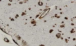
Several neuroimaging parameters have been proposed for diagnosing CAA in vivo. The most commonly used criteria are formulated in the 'Boston' criteria (see Table 1). The detection of large intraparenchymal hemorrhages on brain CT and MRI, in addition to small punctate hemorrhages (so called 'microbleeds', see Figure 1) or superficial collections of blood breakdown products within the brain grooves (so called 'superficial siderosis') detected on MRI, can be suggestive – but is not the final – proof for CAA . Besides, these abnormal findings on neuroimaging represent only the very late stages of CAA. They should be regarded as the 'tip of the iceberg' of the true extent of CAA that is present in the brain.Body fluid biomarkers
Thus, novel biomarkers are needed to accurately detect CAA. Cerebrospinal fluid (CSF) biomarkers may be an excellent candidate for the improved and early diagnosis of CAA, since CSF reflects the brain metabolic state and thus has the ability to reveal pathophysiological changes. For example, earlier work has demonstrated that the concentrations of two isoforms of amyloid β, i.e. amyloid β40 and amyloid β42, in CSF from CAA patients were decreased relative to controls and patients with Alzheimer’s disease.* In addition, we aim to find serum biomarkers.Our research projects
In several research projects, we aim to develop and validate body fluid biomarkers to provide diagnostic tools to clinicians that will allow them to detect CAA during life, to establish the contribution of CAA to cognitive decline and dementia, and to facilitate potential future personalized therapy. Currently active research project comprise:- BIONIC: BIOmarkers for cogNitive Impairment due to Cerebral amyloid angiopathy
- SCALA: An Amyloid Beta Profile Specific for Cerebral Amyloid Angiopathy
- CAFE: Cerebral amyloid angiopathy fluid biomarkers evaluation

Background Cerebral Amyloid Angiopathy
What is CAA? How does it develop? And how can CAA be detected? read moreStart and end date
- Start date: 1 April 2018
- End date: 1 April 2022
The projects

BIONIC project
Diagnosis of cerebral amyloid angiopathy (CAA) currently relies on the 'Boston criteria', which are based on MRI markers. However, these criteria are based on late and indirect hemorrhagic markers of CAA and do not provide definitive proof of the disease; this hampers early diagnosis. read more
CAFE project
Recently, a transgenic rat model of CAA was developed that closely resembles human CAA deposition in the microvasculature. This model allows for studying disease progression in relation to biomarker levels longitudinally, and gives the opportunity to investigate prodromal stages of disease. read more
SCALA project
This project aims to identify a profile of amyloid β-peptides in cerebrospinal fluid that is specific for CAA. If we manage to establish a CAA-specific profile of amyloid β peptide, this offers new possibilities for (early) diagnosis of CAA and monitoring the progression of the disease. read moreGrant providers


Team


