12 October 2018
The researchers, including Peter Friedl and Eleonora Dondossola, the University of Texas MD Anderson Cancer Center, Houston, had been working to develop a better method for studying bone metastases in animal models of cancer for the last six years. A fluorescent imaging technique called intravital multiphoton microscopy (iMPM) has been used extensively to visualize cancer in soft tissues. However, in order to study bone metastases, the researchers had to figure out a way to enable iMPM to look inside much harder material.
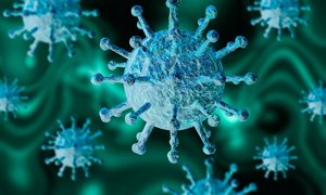
 In this 3D cross-section, you see the nuclei (green) and cytoplasm (red) of human prostate cancer cells growing inside a bioengineered construct of mouse bone (blue-green) that’s been placed in a mouse.
The researchers, including Peter Friedl and Eleonora Dondossola, the University of Texas MD Anderson Cancer Center, Houston, had been working to develop a better method for studying bone metastases in animal models of cancer for the last six years. A fluorescent imaging technique called intravital multiphoton microscopy (iMPM) has been used extensively to visualize cancer in soft tissues. However, in order to study bone metastases, the researchers had to figure out a way to enable iMPM to look inside much harder material.
Related news items
More variants TLR7 gene found in young healthy men with severe Covid-19 Screening and accelerated vaccination for TLR7 immunodeficiency
3 August 2021 Spanish-Dutch research has revealed two new mutations in the TLR7 gene in healthy young men who became seriously ill with severe Covid-19. go to page
Sonlicromanol seems promising for certain cancers Khondrion announces publication in PLOS ONE of new research
13 July 2021 Khondrion announces publication in PLOS ONE of new research showing normalisation of prostate cancer stem cell mPGES-1 overexpression and inhibition of cancer spheroid growth by sonlicromanol’s active metabolite. go to page
Current overview of intriguing RNA therapies
8 April 2021 In an article in EMBO Molecular Medicine, researchers from Radboudumc, together with European colleagues, provide an up-to-date overview of the (im)possibilities of these intriguing RNA therapies. go to page
First online FIJI course
12 June 2020 Although Jack Fransen, Marieke Willemse and Merijn van Erp missed the personal classroom interactions, they can again look back to a successful course thanks to an enthusiastic group of 14 students who helped them through this first online version. go to page
NWO open call GROOT grant for Peter Friedl
24 February 2020 Peter Friedl, theme Cancer development and immune defense, received a 300,000 EUR grant within the NWO-GROOT consortium "Active matter of cancer metastasis" to identify the mechanisms of collective metastasis in breast cancer. go to page