Tom Scheenen has been appointed Professor of Biomedical Magnetic Resonance at the Radboudumc / Radboud University as of October 1st 2021. He aims to bridge the gap between the development of magnetic resonance (MR) techniques and the implementation of new applications in the clinic, improving visualization of cancer. Next to investigating the origin and spread of cancer, he also characterizes the tumor at hand.
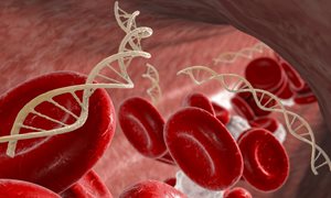
Magnetic resonance imaging (MRI) and spectroscopy (MRS) are versatile techniques that enable non-invasive three-dimensional imaging of patients. These techniques work with a strong magnetic field, which measures the presence and microscopic environment of protons in water, fat and metabolites, providing images with different contrasts of multiple tissues. In this way, cancer can be detected and staged locally, aggressiveness and metastatic spread can be assessed as well as the tumor’s response to treatment.
As a biophysicist, Tom Scheenen focuses on the technical development of MRI and MRS: "Working in the Department of Medical Imaging, I am in direct contact with various medical specialists who can indicate clinical needs. What morphology and which processes in the body are important to a doctor to be visualized? I can choose the best technique and develop it into a clinical application. I’d also like to contribute to more fundamental research: how does a tumor develop in interaction with its surrounding tissue? With MRI, you can do all kinds of measurements on the structure and metabolism of a tumor."
Irono xide nanoparticles
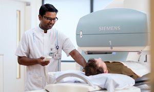
Scheenen works primarily on prostate cancer. In addition to the clinical MRI scanners at the Radboudumc, he also uses an experimental system located at the Erwin L Hahn Institute in Essen. "The new scanner in Essen has a stronger magnetic field than the clinical scanners at the Radboudumc, as much as 7 Tesla. This improves sensitivity and provides a higher spatial resolution of the images," Scheenen explains. In prostate cancer, he uses ultra-small particles of iron oxide (USPIOs) as a contrast agent to look for suspicious lymph nodes that may contain tumor cells. He now also uses this technique for cancer in other parts of the body like the pancreas, esophagus, rectum and head and neck.
Major milestones have been reached in the field of prostate cancer. Thanks in part to Scheenen's research, the so-called multi-parametric MRI (mp-MRI) has been incorporated into (inter-)national clinical guidelines as a first step to detect cancer when prostate-specific antigen (PSA) levels in the blood are elevated, resulting in fewer biopsies. "One next step is the further development of mp-MRI, so that we can reliably detect or predict the presence of small metastases. In addition to anatomy, we also look at blood flow and the mobility of water, which is a measure of the density of tissue. The denser the tissue, the more aggressive the tumor. MRI can be made more quantitative and we are measuring more and more in 3D."
Measuring aggressiveness
In addition to protons in water and fat, Tom Scheenen also measures other atomic nuclei with MRI, such as phosphorus, fluorine, carbon and deuterium. "The concentration of substances with these nuclei is often different in tumors than in healthy tissue. We are looking for a link between these so-called metabolites and the aggressiveness of a tumor, so their presence can help predicting cancer stage and prognosis." That in turn presents new technical challenges. "As soon as we want to measure nuclei other than hydrogen, we need a different measuring device in the MRI scanner. We develop those special parts for our preclinical scanners ourselves, and for clinical use we collaborate with companies like Tesla Dynamic Coils and Rapid Biomedical."
In addition to the projects with companies, Tom Scheenen also works a lot with other research groups. "Magnetic resonance is a broad topic, which interfaces with many other fields at the university and in the hospital. For instance, I am currently working on a project with the Department of Neurology on spectroscopy of the brain. Collaboration is always very welcome, both on our preclinical and clinical scanners, and both inside and outside the Radboudumc."
Career
Tom Scheenen (1974, Roggel) graduated cum laude in Molecular Sciences, with a specialization in Biophysics. He received his PhD from Wageningen University on his thesis titled 'Nuclear Magnetic Resonance Imaging of Water Motion in Plants'. Since 2001, he has been working at the Department of Medical Imaging of the Radboudumc, with a six-month visit at the National Institutes of Health in the United States. Among others, he received a VENI and several ERC grants. Since 2006, he has been working one day a week at the Erwin L Hahn Institute in Essen.
-
Want to know more about these subjects? Click on the buttons below for more news.
More information
Annemarie Eek

wetenschapsvoorlichter