 A computer program that analyzes MRI images can reliably map the presence and even the aggressiveness of a prostate tumor. This is what Radboudumc researchers and international colleagues have published in the scientific journal Investigative Radiology.
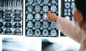
Let’s suppose you’re a man, and you have had elevated PSA levels (prostate-specific antigen) for some time. This might indicate a prostate tumor. A so-called multi-parametric MRI scan would seem advisable. Those MRI images are assessed by a radiologist and given a PI-RADS *) score. Is there indeed a tumor present? If so, how aggressive is it? To determine this, a biopsy is usually performed, in which a small piece of tumor tissue is removed via a hollow needle.
The assessment of the different MRI images is done by humans. It requires a lot of experience to read these images as thoroughly and unambiguously as possible. Years ago, Tom Scheenen and his colleagues from the Radiology and Nuclear Medicine department wondered whether the information from these MRI images could also be assessed automatically through an objective calculation. The idea was to simply circle an anatomically suspect spot in the prostate on the images and then have the machine calculate whether the tissue is healthy or abnormal. And if so, have it objectively determine the severity and diagnose it.
Is it a tumor?
In Investigative Radiology, Scheenen and colleagues have now published research that shows that it is possible. In five different hospitals, MRI images were taken of a total of fifty patients based on a clear protocol. The patients followed the normal examination and treatment route, but it was also examined whether a proper analysis and diagnosis could be made on the basis of just the MRI images.
Researcher Tom Scheenen: “The MRI images of approximately half an hour long provide an enormous amount of information. Firstly, we can distinguish between normal tissue and abnormal tumor tissue. Secondly, we visualize the mobility of water in the abnormal tissue. We also look at how quickly contrast fluid leaks from the blood vessels in and around the tumor, and how it flows back and is absorbed.”
Flows and leaks
The latter two criteria provide information about the severity and aggressiveness of the tumor. Scheenen: "In tumor tissue, cells are packed closer together, and the normal structure between the cells is disturbed. This ensures that water in tumor tissue often encounters boundaries that it is less likely to pass through. The less mobile the water, the more aggressive the tumor. This can be easily visualized with so-called diffusion-weighted MRI, and captured in objective numbers.”
The leakage of contrast fluid from small blood vessels around the tumor is also an important criterion. Scheenen: "A tumor needs extra nutrition and oxygen to grow. To do this, it quickly but carelessly creates new blood vessels. The poor quality of these vessels causes the leakage of the contrast fluid, something that is not the case with normal blood vessels. In these case, the sooner the contrast medium leaks - and is then absorbed and removed - the more aggressive and dangerous the tumor.”
Objective calculation
The MRI images not only make it possible to determine the presence of a prostate tumor but also to assess its severity and aggressiveness objectively. Moreover, the research they have now published shows that the results of the 50 patients in all five participating hospitals could have been assessed in the same way and would have led to the same results. Scheenen: “Our research shows that this approach works and that it is a valid, generally applicable method.” What are the benefits? “Any differences in assessment between radiologists can be prevented because we now have a technique to standardize and automate the assessment. Furthermore, in people with a tumor that is barely growing, checks can now be carried out with an MRI and without a biopsy.” To further investigate the power of the system, Scheenen is entering these and future data into DRE, the Digital Research Environment. "This way, the system will be available to radiologists all over the world, and we will probably be able to improve it even more, adding more data.”
Publication in Investigative Radiology: A Single-Arm, Multicenter Validation Study of Prostate Cancer Localization and Aggressiveness With a Quantitative Multiparametric Magnetic Resonance Imaging Approach - Marnix C. Maas, Geert J.S. Litjens, Alan J. Wright, Ulrike I. Attenberger, Masoom A. Haider, Thomas H. Helbich, Berthold Kiefer, Katarzyna J. Macura, Daniel J.A. Margolis, Anwar R. Padhani, Kirsten M. Selnæs, Geert M. Villeirs, Jurgen J. Fütterer, Tom W.J. Scheenen
*) PI-RADS scale for prostate MRI imaging:
The prostate MRI is assessed on a scale of 1 to 5. The scales indicate the risk of (clinically significant) cancer
- PI-RADS 1: very low (clinically significant cancer is highly unlikely to be present)
- PI-RADS 2: low (clinically significant cancer is unlikely to be present)
- PI-RADS 3: intermediate (the presence of clinically significant cancer is equivocal)
- PI-RADS 4: high (clinically significant cancer is likely to be present)
- PI-RADS 5: very high (clinically significant cancer is highly likely to be present)
Related news items
Millions of euros for study of laser treatment for glioblastoma
2 April 2021 A research group from Radboudumc and UMC Utrecht is to investigate laser treatment of a rare brain tumor, glioblastoma. 3.9 million euros has been made available for the research by Zorginstituut Nederland and ZonMw, under the auspices of the Subsidy Scheme for Promising Care. go to page
Jurgen Fütterer appointed Professor of Image-Guided Oncological Interventions
26 February 2020RIHS researcher Fütterer is an interventional radiologist and an expert in the field of cancer imaging techniques, image-guided interventions and robotics. He is also Professor at the Robotics and Mechatronics Group at the University of Twente.
go to page
SPL Medical starts clinical trial with contrast agent ferrotran
11 February 2020 SPL Medical is starting an international registration study for its contrast agent Ferrotran. The study is being conducted in ten hospitals in Germany, Switzerland and the Netherlands. RIHS researcher Jelle Barentsz discovered the value of this drug in patients with lymph node metastases. go to page
New Guidelines for the detection of prostate cancer MRI instead of tissue samples
3 February 2020 The module on diagnostic prostate MRI for the NVU guidelines for prostate carcinoma took effect on 29 January 2020. The development of these guidelines is due in part to the efforts of RIHS researcher Jelle Barentsz and Ivo Schoots (Erasmus MC). go to page
Jelle Barentsz's trilogy of educational papers in European Urology
12 December 2019 Recently, a trilogy of educational papers by our full professor Jelle Barentsz has been published in European Urology. The publications address what urologists need to know about prostate-MRI. go to page