About the MRI-guided biopsy of the prostate
You have already had an MRI scan (Magnetic Resonance Imaging) of your prostate gland whereby some irregularities have shown up. To be able to determine what exactly these irregularities are, your specialist (Urologist/Radiotherapist) has requested a second MRI examination. read moreAbout the MRI-guided biopsy of the prostate
You have already had an MRI scan (Magnetic Resonance Imaging) of your prostate gland whereby some irregularities have shown up. To be able to determine what exactly these irregularities are, your specialist (Urologist/Radiotherapist) has requested a second MRI examination. During this second examination, visible
abnormalities can be located and small samples can be taken for further analysis by a pathologist (biopsy). This folder will explain the procedure.
On this website, how the examination is carried out is only a general description of a procedure. It may be that your specialist requests a procedure that may vary from the one described here, as it is not possible to list every variant of the procedures that are carried out. Risks and side-effects are only explained in general terms and possible complications will be explained by your specialist.
About the research
Preparation
Unless otherwise requested by your specialist, you may continue to take any medications and eat and drink normally. Rings and piercings made of gold or silver may be worn as the magnet doesn’t affect these items. read morePreparation
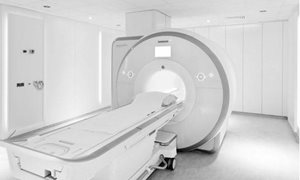
What is an MRI examination?
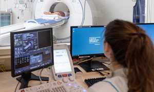
An MRI scan uses a strong magnetic field to create images, of tissues, organs and other structures inside your body. The MRI scanner is like a tube about 1.5 meters long, surrounded by a large circular magnet. You lie on a couch which then slides into the scanner. The part of your body being examined is positioned in the middle of the tube (to obtain the best images) and while each ‘picture’ is being taken you will need to keep still,
otherwise the scan images may be blurred. The examination is carried out by a radiographer together with a radiologist. During your scan the radiographer can see you via a television screen and you will be given a ‘rubber ball’ to hold, which is an alarm signal to the radiographer that you wish to speak to them. MRI has been shown to be extremely safe as long as proper safety precautions are taken. In general, the MRI procedure produces no pain and causes no known short-term or long-term tissue damage of any kind.
Metal /MRI safety
Metal objects in or outside your body are attracted to the strong magnet in the scanner. Therefore, you may not enter the MRI examination room with, for example, a wheelchair, keys or coins. Credit/bank cards with a magnetic chip, hearing aids, mobile phones and watches, are damaged when close to the strong
magnet in the scanner (the magnet is always switched on!).
Should you (or your companion) have a pacemaker/defibrillator (ICD), a neurostimulator in the spine, aneurysm clips, dentures/teeth with implanted magnetic fastenings, a bladder stimulator or a permanent insulin pump, then you may not enter the examination room. This also applies to foreign metal objects, such as metal splinters (especially in or near the eyes), shrapnel or bullet wounds and older types of heart valves, aneurysm clips or certain cochlear (ear) implants. Modern prosthetics, such as hip or knee replacements, are not usually a problem.
We request that you complete the attached ‘checklist’ and bring it with you to your appointment. Should you have answered any questions with a ‘yes’, we request that you contact the Radiology Department.
How to Prepare for the MRI Examination
Unless otherwise requested by your specialist, you may continue to take any medications and eat and drink normally. Rings and piercings made of gold or silver may be worn as the magnet doesn’t affect these items. All other items of jewellery and watches are best left at home. You will be given a gown to wear.
- Antibiotics
Your specialist will prescribe a short course of antibiotics in the form of 2 tablets of Ciproxin® 500 mg. These tablets should be taken orally. You need to take 1 tablet 2 hours before the biopsy procedure and 1 tablet 10 hours after the biopsy procedure. If you have a known allergy to antibiotics or have had an earlier biopsy whereby you later developed an infection, it is important to inform our department and your specialist of this.
- Anticoagulant Therapy (blood-thinning medication)
When you take anticoagulants inform your specialist. You should discuss with your specialist as to when you need to stop taking these medications prior to your biopsy. This should be noted on the biopsy request form. You must also inform your thrombosis service that your INR must be 2 or lower on the day of the examination and the reason being is that you are undergoing a biopsy on that day. You are expected yourself to make an appointment with the thrombosis service to have your INR measured. Please bring with you the written results of this blood test. If your blood clotting time is too long, then the procedure cannot go ahead due to an increased risk of haemorrhage.
When you take Dabigatran or Rivaroxaban, then there is no need to check your coagulation status. These medicines also need to be reported to your doctor. Your doctor will let you know how many days before the biopsy you need to stop the intake of this medication. Your doctor will decide about this based on your kidney
function.
- Medications
Usually you can continue to take your medications as prescribed, however your doctor may advise you to stop temporarily taking certain medications. We would advise you to bring your medication passport (available from your pharmacist) or alternatively, make a list of all medications that you are taking at the time of your scan.
- Heart Defects
Do you have any of the following conditions:
• A history of endocarditis? (an inflammation of the lining of the heart)
• Defects, congenital or otherwise, of your heart, heart valves or the great vessels of the heart?
• Cardiomyopathy? (a disease of the heart muscle)
• Foreign materials, clips, stents or implants in your heart or the great vessels of your heart?
If the answer to any of these questions is ‘yes’, we request that you inform our department or your specialist as soon as possible as you may require an extra course of antibiotics.
- Claustrophobia
People with severe claustrophobia may be anxious about undergoing an MRI scan. Should you have severe claustrophobia, then please inform your specialist or GP of this, as they may prescribe a mild sedative. Even a mild sedative will affect your ability to drive, so please make sure that you make other arrangements for transportation to the hospital.
The Examination
You are required to report to the Radiology Department (Route 780) 10 minutes before the time stated on your appointment card or letter, keeping in mind that it’s a 10 minute walk from the main entrance to the Radiology Department. Should you require assistance with mobility, transportation can be arranged at the reception desk in the main hall. When you check in at the Radiology Department, your details will be verified and corrected if necessary. You will then be directed to the correct waiting area. Should you have forgotten to bring your ´check list´ then you will be required to fill out a new questionnaire. You will then be directed to the correct waiting area. The radiographer or assistant will collect you (and your companion) from the waiting area, bring you to a changing room where you (and your companion) will be required to leave all metal objects, telephones, credit-bank cards etc, behind. You can put them in a safe.
During the procedure
You are asked to arrive at the Radiology department (route 780) 10 minutes prior to the appointment time. A radiology employee will take you to the preparation room and explains the procedure. read moreDuring the procedure
You are asked to arrive at the Radiology department (route 780) 10 minutes prior to the appointment time. A radiology employee will take you to the preparation room and will explain the procedure. You are asked to go to the toilet and to change your clothes for a surgical gowns and pants. Hereafter, he radiology employee will bring you in a wheelchair to the MR room at the OR complex. There you are asked to lie down on your back on the scanner table.An antenna in the form of a plate (also used in the previous MRI scans) will be placed over your pelvis. Your pelvis is covered with sterile drapings and the skin between your anus and scrotum will be locally anaesthetized with an injection. Then, several images of your prostate are acquired to determine the exact location for biopsy. The physician will insert the needle in the skin between your anus and scrotum. Under MR image guidance it will be inserted into the prostate. This needs to happen stepwise, because we have to be very accurate. Once the physician is convinced that the needle is in the correct position, the biopsy will be performed. Acquiring these samples may be uncomfortable. A control scan is made, to check the position of the needle. In general, 3 to 4 samples are taken from each suspicious area. The whole procedure takes 60-90 minutes.
After care
After your examination you cannot participate in traffic. We recommend to bring someone with you to drive you home, or to make use of public transport. read moreAfter care
After your examination you cannot participate in traffic. We recommend to bring someone with you to drive you home, or to make use of public transport. You may eat and drink normally after your examination. After a biopsy you may have some bleeding from the rectum, blood in your stools (up to 2 days post-biopsy), blood in your urine (up to 4 days post-biopsy) or blood in your semen (up to 30 days post-biopsy). If you develop a fever (temperature above 38.5°C), have cold shivers, have abdominal pains or feel ill, then you should immediately contact the nearest emergency department, as this can be an indication of an inflamed prostate gland. In this case you should inform the physician that you have had an endo-rectal biopsy. We would like to be informed should you develop any complications due to this procedure. We would appreciate it if you would contact us via (024) 365 22 79 or (024) 365 22 82. This number is available only on weekdays between 08.15-16.15The results
The results of your biopsy will not be available immediately. Within 2 weeks of the procedure, your specialist will receive the results.More information
Your appointment
Your doctor will make an appointment. You will get a notification. If you are unable to attend, please let us know as soon as possible so that your appointment can be given to another patient.
lees meerYour appointment
Your doctor will make an appointment. You will get a notification.
Cancelling or changing your appointment
If you are unable to attend, please let us know as soon as possible so that your appointment can be given to another patient. You can call the Radiology Department on weekdays between 08.30-16.45 at (024) 361 45 29.
Should you wish to cancel your appointment we request that you also inform your specialist.
Getting there
Visiting address
Radboudumc main entrance
Geert Grooteplein Zuid 10
6525 GA Nijmegen
Directions


Department Medical imaging
The department of medical imaging examines and treats various conditions, and has three focus areas: anatomy, nuclear medicine and radiology.
go to page