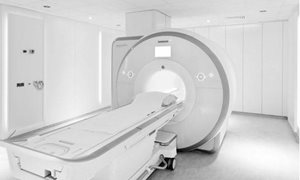
Why an MRI-scan?
An MRI scan uses a strong magnetic field and radio waves (not X-rays) to create pictures, on a computer, of tissues, organs and other structures inside your body. An MRI scan uses a strong magnetic field and radio waves (not X-rays) to create pictures, on a computer, of tissues, organs and other structures inside your body.MRI Checklist
Please read and complete the following checklist before your appointment. read moreThe examination
Before the examination
Before the examination, some precautions should be taken. Moreover, some preparations should be taken care of.
read moreBefore the examination
Metal
Metal objects in or outside your body are attracted to the strong magnet in the scanner, therefore you may not enter the MRI examination room with, for example, a wheelchair, keys or coins. Credit/bank cards with a magnetic chip, hearing aids, mobile phones and watches are damaged when close to the strong magnet in the
scanner (the magnet is always switched on!).
Should you (or your companion) have a pacemaker/defibrillator (ICD), a neurostimulator in the spine, aneurysm clips, dentures/teeth with implanted magnetic fastenings, a bladder stimulator or a permanent insulin pump, then you may not enter the examination room. This also applies to foreign metal objects, such as metal splinters (especially in or near the eyes), shrapnel or bullet wounds and older types of heart valves, aneurysm clips or certain cochlear (ear) implants. You may be required to remove medication patches if they contain metal foil. Usually modern hip and knee replacements are not a problem.
Clothing and jewelry
Unless otherwise requested by your specialist, you may continue to take any medications and eat and drink normally. Piercings made of gold or silver may be worn as the magnet doesn’t affect these items. All other items of jewellery and watches are best left at home. Please wear comfortable clothing with separate top and bottoms. Should any clothing contain metal parts (e.g. zips or underwired bras) you will be given a gown
to wear. Please don’t apply hairspray and make-up as they can affect MRI images and cause some irritation.
Medications
Usually you can continue to take your medications as prescribed, however your doctor may advise you to temporarily stop taking certain medications. We would advise you to bring your medication passport (available from your pharmacist) or alternatively, make a list of all medications that you are taking at the
time of your scan.
Pregnancy and breast-feeding
There is no evidence to suggest that MRI scans pose a risk during pregnancy. However, as a precaution, scanning is not usually recommended during the first 12 weeks of pregnancy. Contrast agents are not used throughout the whole term of the pregnancy. Should you be or think that you may be pregnant, please contact the Radiology Department.
Only a limited amount of contrast agent enters breast milk. Therefore, it is safe to breastfeed. If you uncomfortable with this, you can express breast milk and discard the milk for 24 hours.
Claustrophobia
People with severe claustrophobia may be anxious about undergoing an MRI scan. Should you have severe claustrophobia, then please inform your specialist or GP of this as they may prescribe a mild sedative. Even a mild sedative will affect your ability to drive, so please make sure that you make other arrangements
for transportation to the hospital.
During the examination
You are required to report to the Radiology Department (Route 780) 10 minutes before the time stated on your appointment card or letter, keeping in mind that it’s a 10 minute walk from the main entrance to the Radiology Department. read moreResults
The results of your MRI scan will not be available immediately. A radiologist will interpret the images and report to your specialist. You will receive the results from your specialist at your next appointment.More information
Getting there
Visiting address
Radboudumc main entrance
Geert Grooteplein Zuid 10
6525 GA Nijmegen
Directions


Changing your appointment
Would you like to change or cancel your appointment? You can indicate this in your online personal medical file ’mijnRadboud’ (only in Dutch) or contact the department where you have an appointment. We will schedule a new appointment with you as soon as possible. login mijnRadboud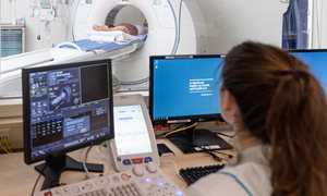
Department Medical imaging
The department of medical imaging examines and treats various conditions, and has three focus areas: anatomy, nuclear medicine and radiology.
go to page