RIMLS highlights 2018
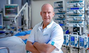
Peter Pickkers Bringing life to the pharmaceutical graveyard (interview)
Infectious diseases and global health read interviewPeter Pickkers Bringing life to the pharmaceutical graveyard (interview)
There aren’t that many researchers that can boast being published in one of the leading medical journals. Less so, as leading principal investigator of that research. Peter Pickkers’ research into sepsis-associated acute kidney injury was published in the October 2018 edition of The Journal of the American Medical Association (JAMA).
Q1 That must feel like a huge accomplishment of your work. How did you feel when your article was accepted?
I knew we had discovered something significant
“It’s the best feeling. By being published by a journal like JAMA, the relevance your research is acknowledged, and you realize the vast effect your results could have for patients. It’s the outcome of a long process.”
“It really started in 2005. In the first phase 2 study with Alkaline Phosphatase, we measured biomarkers in urine as an aside thought. The results were really interesting. A new small trial was designed aimed to confirm the kidney protective findings, never expecting to reproduce the same results. When we did, I knew we had discovered something significant, and the focus completely shifted to the kidney. It started with luck – so to say.
“Sepsis is the leading cause of death worldwide. Research into sepsis is known as the pharmaceutical graveyard and clinical research related to renal failure has a similar track record. Decades of research and dozens of projects have not led to a single, effective drug. Getting funding in such a field during the financial crisis was not easy for the company."
Q2 Can you tell me a bit about the research and results that you published in JAMA?
“The article in JAMA is the result of a three-year project, looking at 301 patients in 53 ICU’s in 11 countries. The objective was to determine the optimal therapeutic dose and effect on kidney function in patients who are critically ill with sepsis-associated AKI. Strictly speaking, the primary endpoint was impact on creatinine clearance during the first seven days of being admitted to the ICU. This didn't result in a significant difference with the placebo. Nevertheless, we also investigated long-term renal function and survival up to 90 days, and found that long-term renal function was better and the mortality rate almost halved in the treatment-group. And these effects were significant.
"The absence of an effect during the first 7 days almost caused us not to get published. There are stringent rules to not report any secondary outcomes in the abstract. I’m glad JAMA recognized the impact of these clinical outcomes."
Q3 You were the primary investigator of the project. Did you feel the pressure to achieve results?
“The most important thing for me is that results are valid. Of course, positive results are great – the cherry on top – but it’s all about quality. I don’t really think about the money of the investors. I want to be completely free to focus on the optimal design. To find something that might be there. I believe, there’s plenty of research with good ideas that has failed due to poor design, especially in the sepsis field.
"On an academic level, this project gave me a great opportunity. My group discovered this effect and I remained involved all the time. Together with the company, we selected the best researchers in the field as national coordinators in their countries. I felt like the ‘young guy’ leading my idols, the experts. Now I can truly say I’m one of them.”
Q4 What does getting published in such a prestigious journal mean for you, the RIMLS institute and, in general, research in this field?
“I’ve been asked to speak at several large conferences, reaching audiences of thousands. It’s good for my reputation but also for RIMLS'. As far as ‘branding’ goes, Nijmegen is definitely known in the intensive care field.
“Of course, friends and family know what this means to me. The barriers I had to overcome to get here. But,” laughing Peter adds, “it’s not like I talk about it at birthday parties.
"Being published in a top-notch general journal offers broad exposure — more than if it's published in a journal about a specific field. More scientists will read it, many from other disciplines. It’s good for the creation of ideas.
“And it’s good for research into sepsis. As I mentioned before, it was considered the pharmaceutical graveyard. I hope it motivates scientists and investors. There’s much more potential.”
Q5 Where do you go from here? What are your hopes for the future?
“All those years ago, we started with an observation in patients, took it back to the lab, deciphered the mechanism of action, and now we’re bringing it back to patients. We’re busy with the design of a phase 3 trial, possibly enrolling up to 1,500 patients in 200 ICU’s all over the world. It’s great to be asked to be the principle investigator again.
“Our objective: an effective drug that limits kidney failure and improves survival of ICU patients with sepsis-associated AKI. If we can confirm this, and the drug will become standard treatment, I feel I’ve made a real impact. That would be a dream come true. But dreams mean work, so let’s get to it!”

Siroon Bekkering et al. Cell
Infectious diseases and global health read moreSiroon Bekkering et al. Cell
Metabolic induction of trained immunity through the mevalonate pathway.
The immune system not only detects and destroys pathogens such as microbes but also plays a role in the onset of diseases such as arteriosclerosis.Scientists from Radboud university medical center, together with fellow scientists from Bonn, Dresden and Pennsylvania, studied a new mechanism that regulates the immune system’s functioning. For example, they discovered that cholesterol inhibitors may prevent infections, that a high-fat Western diet can have a long-lasting effect on our immunity and that even our stem cells can be disrupted. Today, four publications on the manipulation of our immune system appear in Cell and Cell Host & Microbe (three articles in Cell, one article in Cell Host & Microbe).
Our immune system consists of two parts: innate immunity and adaptive immunity. Both help in fighting diseases but there is one big difference. Our innate immune system works rapidly and non-specifically: it destroys all invading organisms. Our adaptive immune system is more accurate: these immune cells are able to distinguish between the body’s own cells and foreign cells. If they encounter an invader such as a microbe, they produce both a specific immune cell and a memory cell. This memory cell reacts rapidly on subsequent exposure to the same organism. Cells of our innate immune system, however, also have some capacity to ‘remember’ things. When they encounter an invader, they may be reprogrammed. If they are subsequently exposed to a random invader, the immune cells will show an enhanced response. This is called ‘trained immunity’. However, this training does not always turn out well. Overactive immune responses can sometimes even cause diseases.
Legend:
Graphical Abstract
Bekkering S., Arts R.J.W., Novakovic B., Stunnenberg H., Riksen N., Netea M.G. Metabolic induction of trained immunity through the mevalonate pathway. Cell 172: 135-146, 2018.
View this publication on NCBI.

Ellen van den Bogaard et al. Genet Med.
Inflammatory diseases read moreEllen van den Bogaard et al. Genet Med.
Deficiency of the human cysteine protease inhibitor cystatin M/E causes hypotrichosis and dry skin.
Their previous genetic studies in mice and in vitro biochemical studies using human enzymes and inhibitors have provided information on the role of the human cysteine protease inhibitor cystatin M/E, encoded by the CTS6 gene, in epidermal cornification and maintenance of hair follicle integrity. Recently they have identified a new autosomal-recessive hypotrichosis syndrome caused by a loss-of-function mutation in the CST6 gene. These findings support the importance of protease-antiprotease balance in the regulation of epidermal and hair follicle homeostasis.
We aimed to assess the biological and clinical significance of the human cysteine protease inhibitor cystatin M/E, encoded by the CTS6 gene, in diseases of human hair and skin.
Exome and Sanger sequencing was performed to reveal the genetic cause in two related patients with hypotrichosis. Immunohistochemical, biophysical, and biochemical measurements were performed on patient skin and 3D-reconstructed skin from patient-derived keratinocytes.
We identified a homozygous variant c.361C>T (p.Gln121*), resulting in a premature stop codon in exon 2 of CST6 associated with hypotrichosis, eczema, blepharitis, photophobia and impaired sweating. Enzyme assays using recombinant mutant cystatin M/E protein, generated by site-directed mutagenesis, revealed that this p.Gln121* variant was unable to inhibit any of its three target proteases (legumain and cathepsins L and V). Three-dimensional protein structure prediction confirmed the disturbance of the protease/inhibitor binding sites of legumain and cathepsins L and V in the p.Gln121* variant.
The herein characterized autosomal recessive hypotrichosis syndrome indicates an important role of human cystatin M/E in epidermal homeostasis and hair follicle morphogenesis.
Legend:
3D visualization of the cystatin M/E–legumain–cathepsin V complex. The visualization was made by superposing Protein Data Bank (PDB) files 4N6O and 3KFQ. Legumain and cathepsin V are shown in purple and magenta respectively. Cystatin M/E is colored by element (β-strands = red, ɑ-helix = blue, loops = cyan/green). The residues deleted after truncation of the protein are colored gray. (a) Overview of the complex in ribbon presentation. (b) Overview of the complex with protein surface shown. (c) Close-up of the protein–protein interaction sites with side chains of the residues shown.
van den Bogaard E.H.J., van Geel M., van Vlijmen-Willems I.M.J.J., Jansen P.A.M., Peppelman M., van Erp P.E.J., Atalay S., Venselaar H., Simon M.E.H., Joosten M., Schalkwijk J., Zeeuwen P.L.J.M. Deficiency of the human cysteine protease inhibitor cystatin M/E causes hypotrichosis and dry skin. Genet Med. doi: 10.1038/s41436-018-0355-3, 2018.
View this publication on NCBI.

Eleonora Dondossola et al. Sci Transl Med.
Cancer development and immune defense read moreEleonora Dondossola et al. Sci Transl Med.
Intravital microscopy of osteolytic progression and therapy response of cancer lesions in the bone.
A system allowing microscopic monitoring and imaging of cancer that has spread to the bone in mice so they can better understand and develop treatment for bone metastasis in humans. The researchers show how the technique can monitor and capture the dynamics of tumor cell interaction with bone and bone resident cells as they occur over time.
- A tissue-engineered construct under a mouse’s skin develops in about a month into bone with an internal cavity and a thin outer layer that the microscope can “see” through.
- After bone marrow and other cells populate the cavity, a cancer cell line is injected.
- Interactions between malignant cells and bone cells are viewed through the multiphoton microscope via a small glass window sewn into the skin above the bone.
Multiphoton microscopy is a fluorescent imaging technique used to image living tissue. The microscopes in the lab of Peter Friedl, can capture up to seven parameters at a time. Friedl is senior author of the study.
The bone construct was developed by a team of scientists at the Centre in Regenerative Medicine, Institute of Health and Biomedical Innovation, Queensland University of Technology, in Brisbane, Australia, led by Dietmar Hutmacher. Normal bone biology involves a balance between bone-creating cells called osteoblasts, and bone-destroying cells called osteoclasts, Eleonora Dondossola, the first author of the article and senior team member in Peter Friedl's laboratory at MD Anderson Cancer Center, notes. Cancer tips this balance, altering the equilibrium between these two populations and leading to symptomatic bone remodeling. The team’s microscopy showed bone loss concentrated around osteoclasts near the tumor. This phenomenon is a known and painful issue for patients with prostate cancer bone metastasis, and a class of drugs called bisphosphonates is used to relieve this symptom. In the clinic, the effect is known to be palliative, relieving pain but not prolonging survival. The team’s multiphoton microscopy captured this effect. They treated the mice with the bisphosphonate zoledronic acid and found that the drug did not reduce the number of osteoclasts, but slowed their activity, preserving bone. Notably, the treatment had no effect on tumor growth, and this explains why the bone is stabilized but patient survival is not prolonged.
Friedl’s lab is using this model to study cancer treatments in mice, including co-clinical work in immunotherapy and radiation. Drugs that free the immune system to attack cancer are often thwarted by resistance factors in the tumor microenvironment, which the team hopes to observe and characterize.
Legend:
PC3 tumor cell administration and monitoring by histology and iMPM. (A) Schematic representation of the model. PC3 cells were injected into the mature ossicle 30 days after implantation into nude mice. A dorsal skinfold chamber (DSFC) was applied 10 to 14 days after tumor cell implantation, adjacent to the ossicle to enable iMPM. (B) Histology of PC3 cells in mTEBC and patient biopsy. T, tumor. Scale bar, 100 μm. (C) iMPM detection of PC3 cells at different scanning depths (z-stack at z = 0, −50, and −96 μm). Overviews shown as xy and xz, yz orthogonal sections, and detail with subcellular resolution (dotted box). Dashed line in top left denoted the edge of the ossicle with fluorescence signal originating from the tumor. (D) XZ intensity profile of individual channels with increasing scanning depth into the ossicle. (E) Quantification of interphase (IF), mitotic, or apoptotic cells in 3D stacks captured by iMPM. Mean ± SD is shown (n = 5 independent lesions). Nucleus, H2B/eGFP (green); cytoplasm, DsRed2 (red); collagen fibers: bone, SHG (cyan). Scale bar, 100 μm.
Dondossola E., Alexander S., Holzapfel B.M., Filippini S., Starbuck M.W., Hoffman R.M., Navone N., De-Juan-Pardo E.M., Logothetis C.J., Hutmacher D.W., Friedl P. Intravital microscopy of osteolytic progression and therapy response of cancer lesions in the bone. Sci Transl Med. 10: eaao5726, 2018.
View this publication on NCBI.

Daniel Garza et al. Nat Microbiol.
Tumors of the digestive tract read moreDaniel Garza et al. Nat Microbiol.
Towards predicting the environmental metabolome from metagenomics with a mechanistic model.
What do the bacteria eat, that live on your skin? Scientists at Radboudumc and Utrecht University have developed a novel computer model to answer this question, revealing that a lot of the food for skin bacteria is derived from beauty and skin care products. The model uses microorganisms’ DNA to determine the metabolites – nutrients and waste products – used and produced by the bacteria in different parts of the human body. Metabolites provide important clues about human health and disease but can be difficult to measure experimentally. The results of their research were published in Nature Microbiology.
Metabolites, such as those that make up one’s body odour, are molecules that are used and produced by living cells such as human cells and bacteria. The human body is increasingly seen as a complex ecosystem, home to thousands of microorganisms and their metabolites. The health of this ecosystem hinges on the relationship between the microbes and human cells and tissues, and metabolites are often the key to this relationship. For example, intestinal tumours cause changes to the metabolite composition in the intestine, which in turn allows certain types of gut bacteria to grow, sometimes exacerbating the tumour.
Difficult to determine
“Metabolites are vital, but it is often difficult to find out which ones are present at a specific site in the body. There are many different metabolites, and their concentrations can vary widely,” explains research leader Dr. Bas Dutilh from Utrecht University. “Since metabolites are so closely linked to the growth of bacteria, we thought that it might be possible to predict their composition based on the types of bacteria present. The only input we need for that is the abundances of the bacteria and their genes, and progress in the field of metagenomics has made it relatively easy to get those.”
Surprisingly consistent
Given gene information from the bacteria as input, the computer model simulates the most likely metabolites that the bacteria need to grow. These are found by checking which enzyme each gene encodes, and which metabolites each enzyme uses/produces. The researchers then calculate the concentrations of metabolites that explain the abundances of bacteria observed at a given site in the human body. “The predictions we arrive at in our analyses are surprisingly consistent with what is known about the metabolite concentrations in and on the human body,” explains lead author and PhD candidate Daniel Garza, affiliated with Radboudumc.
“We have shown the utility of the computer model for the human skin, mouth, intestines and vagina, because those are fairly well-known areas,” says Prof. Martijn Huijnen from Radboudumc. “But in principle, this technique is applicable to any environment where bacteria live, such as the soil near plant roots.”
Beauty and skin care products
The researchers found that many of the substances predicted from skin samples are known to be ingredients of beauty and skin care products, such as myristic acid, a fragrance ingredient that is easily absorbed by the skin. The results also predicted a significant amount of lead on skin, which corresponds to the findings of a previous experimental study. Where the toxic metal comes from, however, is still anyone’s guess. Lead supplement in petrol has long been prohibited in the Netherlands and the United States, where the samples were taken. “It’s still used in some brands of lipstick, but that doesn’t seem to be a likely explanation for the relatively high concentrations found in so many skin samples”, according to Dutilh.
The model in detail
The computer algorithm, called MAMBO (for ‘Metabolomic Analysis of Metagenomes using fBa and Optimization’), works as follows (see figure). (1) First, computer models are created of the metabolisms of more than 1,500 bacteria from the human body. These models record the relationship between the concentration of metabolites in the environment and the growth of each bacteria. (2) The models are used to conduct computer simulations of bacterial growth on an initially random, but well-defined metabolite environment. This produces a predicted profile indicating which bacteria will grow in the selected metabolite environment, and how well they grow. (3) The predicted profile is then compared to an experimental profile, for example one listing the abundances of all the bacteria found in samples from skin or faeces. (4) After simulating millions and millions of environments, the predicted profile that best corresponds to the experimental profile is found. This prediction describes all the bacteria and metabolites that are found and at what concentrations. Thus, the metabolite concentrations in an environment can be predicted by measuring the bacterial genes.
Legend:
Overview of the MAMBO algorithm. Reference microbial genomes are used to reconstruct GSMMs (1); community abundance profiles are obtained through reference mapping (2); and biomass production of the GSMMs, obtained through FBA (3), is correlated with the metagenomic community abundance profile (4). This correlation is optimized by multiple iterations of a Monte Carlo-based sampling algorithm.
Garza D.R., van Verk M.C., Huynen M.A., Dutilh B.E. Towards predicting the environmental metabolome from metagenomics with a mechanistic model. Nat Microbiol. 3:456-460, 2018.
View this publication on NCBI.

Jakob Goldmann et al. Nat Genet
Mitochondrial diseases read moreJakob Goldmann et al. Nat Genet
Germline de novo mutation clusters arise during oocyte aging in genomic regions with high double-strand-break incidence.
Clustering of mutations has been observed in cancer genomes as well as for germline de novo mutations (DNMs). We identified 1,796 clustered DNMs (cDNMs) within whole-genome-sequencing data from 1,291 parent–offspring trios to investigate their patterns and infer a mutational mechanism. We found that the number of clusters on the maternal allele was positively correlated with maternal age and that these clusters consisted of more individual mutations with larger intermutational distances than those of paternal clusters. More than 50% of maternal clusters were located on chromosomes 8, 9 and 16, in previously identified regions with accelerated maternal mutation rates. Maternal clusters in these regions showed a distinct mutation signature characterized by C>G transversions. Finally, we found that maternal clusters were associated with processes involving double-strand-breaks (DSBs), such as meiotic gene conversions and de novo deletion events. This result suggested accumulation of DSB-induced mutations throughout oocyte aging as the mechanism underlying the formation of maternal mutation clusters.
Legend:
(a) Z scores of expected and observed overlaps of cDNM clusters in our cohort and sex-matched meiotic gene conversion in another cohort21. Diamonds, observed values. Box plots: box, interquartile range; line, median; whiskers, extreme values >1.5× interquartile ranges from box borders. (b) DNMs detected close to sites of de novo CNVs. Data for DNMs are listed in Supplementary Table 15. (c) cSNP density close to CNV breakpoints (Methods). (d) Z scores of expected and observed overlap of cDNM clusters and sex-matched recombination hotspots. Symbols and box plots as in a.
Goldmann J.M., Seplyarskiy V.B., Wong W.S.W., Vilboux T., Neerincx P.B., Bodian D.L., Solomon B.D., Veltman J.A., Deeken J.F., Gilissen C., Niederhuber J.E. Germline de novo mutation clusters arise during oocyte aging in genomic regions with high double-strand-break incidence. Nat Genet. 50:487-492, 2018.
View this publication on NCBI.

Arjan van Laarhoven et al. Lancet Infect Dis.
Infectious diseases and global health read moreArjan van Laarhoven et al. Lancet Infect Dis.
A completely new insight regarding the high mortality of TB meningitis.
Meningitis is the most severe manifestation of tuberculosis (TB), leading to death or disability in at least 50% of those affected. Diagnosis is difficult, and optimal antibiotic treatment has not been defined. TB meningitis is relatively rare, with approximately 20 cases per year in the Netherlands. Reinout van Crevel, professor in Global Health and Infectious Diseases, has worked on TB meningitis with colleagues at Padjadjaran University / Hasan Sadikin hospital in Bandung, Indonesia for over 10 years. In previous studies van Crevel and colleagues have focused on diagnosis of TB meningitis and shown that intensified antibiotic treatment with a higher dose rifampicin reduces mortality of TB meningitis (Ruslami et al., Lancet Inf Dis 2013).
Immunopathology contributes to the high mortality of tuberculous meningitis, but it is mostly unknown which biological pathways are involved, and corticosteroids are only effective in reducing mortality in some patients. As part of his PhD studies, Arjan van Laarhoven (PhD) has used the established Indonesian cohort and bioarchive of over 800 patients with TB meningitis to identify what pathways are involved in poor outcome of TB meningitis.
More than 400 metabolites were measured in archived CSF and serum samples from a discovery set of patients. Most CSF metabolites levels were higher in TBM patients, especially in those who died during follow-up. CSF tryptophan concentrations showed a different pattern: concentrations were much lower in patients who survived compared to patients who died (9-fold) and to controls (31-fold). The association of low CSF tryptophan with patient survival was confirmed in the second cohort of over 100 patients. Finally, genome-wide SNP typing was used to identify tryptophan quantitative trait loci (QTLs), and these same loci strongly predicted mortality in a third analysis in a third cohort of 285 TB meningitis patients.
These findings clearly demonstrate that cerebral metabolism of tryptophan, an essential amino acid which is known to affect Mycobacterium tuberculosis growth and central nervous system inflammation, is critical for the outcome of TB meningitis.
This study is important for three reasons. First, it has led to a real new insight in the pathways that may be involved in the immunopathology and death of tuberculous meningitis, the most dramatic and lethal manifestation of tuberculosis. These findings are a first step towards more effective, and possibly personalised host-directed therapy in TB meningitis, and possibly in other brain infections. Second, this is the study first that links metabolomics and genome-wide genotyping with mortality of tuberculosis in a 'systems approach’. This expertise builds on the functional genomics work that Mihai Netea from the department of Internal Medicine has established with many collaborators in- and outside Radboudumc. Third, this study is evidence of the scientific and clinical impact of long-term collaboration in infectious disease research with partners in Indonesia and other tropical countries.
Legend:
CSF metabolome as a determinant of survival in patients with tuberculous meningitis. (A) Individual metabolites in CSF with ratio between tuberculous meningitis survivors and non-survivors, and ratio between tuberculous meningitis survivors and controls. Colours indicate strength of association; metabolites that do not show differences between groups (uncorrected p>0·05) are grey. The three subplots show metabolite concentrations according to patient category. The y axis shows the 2-log of the relative abundance of metabolite ions as chromatographic peaks (peak ion intensity). Leukotriene B4, glucose, and tryptophan were chosen as relevant metabolites representing three different quadrants of the plot. (B) Kaplan-Meier plot of patient survival in the tryptophan validation cohort, according to CSF tryptophan concentrations, as divided in the following tertiles: low (<0·18 μmol/L), intermediate (0·18–0·69 μmol/L), and high (>0·69 μmol/L). CSF=cerebrospinal fluid.
van Laarhoven A, Dian S, Aguirre-Gamboa R, Avila-Pacheco J, Ricaño-Ponce I, Ruesen C, Annisa J, Koeken VACM, Chaidir L, Li Y, Achmad TH, Joosten LAB, Notebaart RA, Ruslami R, Netea MG, Verbeek MM, Alisjahbana B, Kumar V, Clish CB, Ganiem AR, van Crevel R. Cerebral tryptophan metabolism and outcome of tuberculous meningitis: an observational cohort study. Lancet Infect Dis. 18: 526-535, 2018.
View this publication on NCBI.


Annemarie Post et al. Clin Cancer Res.
Women's cancers read moreAnnemarie Post et al. Clin Cancer Res.
Common features of tamoxifen resistance and radioresistance in breast cancer.
Treatment resistance is a main cause of adverse disease outcome in breast cancer patients. Therefore, this study aimed to identify common features of tamoxifen- and radioresistance in breast cancer cells in vitro and investigate relevant pathways in patient cohorts to establish the relevance of the identified pathways.
Both tamoxifen- and radioresistant breast cancer cells had increased expression levels of genes involved in type I interferon signaling, compared to non-resistant cells. IFN-stimulated genes (ISGs) were induced in a dose-dependent and time-dependent manner after tamoxifen treatment and irradiation. Tamoxifen treatment also led to ssDNA presence in the cytoplasm, a phenomenon that has already been described for irradiation and is known to induce expression of ISGs.
Several breast cancer patient cohorts were analyzed to investigate the relation of ISG expression with treatment sensitivity. In one cohort, high expression levels of ISGs were found in the primary tumor in around half of the patients. This was associated with a tumor infiltrating lymphocyte expression signature, while the ISGs were also expressed by the tumor cells themselves. Importantly, the expression of ISGs correlated to outcome in breast cancer patients treated with adjuvant tamoxifen or radiotherapy, but not in systemically untreated patients or chemotherapy-treated patients.
These data indicate that expression of ISGs by tumor cells is involved in acquired, treatment-induced resistance to tamoxifen and radiotherapy, and might play a role in intrinsic resistance via interaction with tumor infiltrating lymphocytes.
Legend:
SGs are coexpressed in breast tumors, and correlate with a TIL expression signature. A, Pearson correlation coefficients between the top 25 of the 94 common genes in MCF-7TAM and MCF-7RT were clustered and visualized (red shows positive correlation, green negative) for a cohort of breast cancer patients. The rectangle indicates genes involved in IFN signaling. B, Heatmap showing relative gene expression of the 18 correlated ISGs (individual probes for each gene on vertical axis; green, levels below median; red, above median) in 155 primary breast cancer patients (on horizontal axis). C and D, Boxplot of STAT1 expression (C) and boxplot of a TIL signature expression (D) in ISGHIGH and ISGLOW patients, showing the median, 25th and 75th percentile. The whiskers extend to the minimum and maximum values. Statistical significance was determined by Mann–Whitney U testing. ***, P < 0.0001.
Post A.E.M., Smid M., Nagelkerke A., Martens J.W.M., Bussink J., Sweep F.C.G., Span P.N. Interferon-Stimulated genes are involved in cross-resistance to radiotherapy in tamoxifen-resistant breast cancer. Clin Cancer Res. 24: 3397-3408, 2018.
View this publication on NCBI.

Liping Qiu et al. Small
Nanomedicine read moreLiping Qiu et al. Small
Endolysosomal-escape nanovaccines through adjuvant-induced tumor antigen assembly for enhanced effector CD8+ T cell activation.
Vaccination is a powerful strategy to induce the activation of tumor‐specific effector immune cells, which is key for successful immunotherapy. However, the generation of effective anticancer nanovaccines is challenging. One of these challenges is the efficient co-encapsulation of antigenic peptides or proteins together with antigen-presenting cell activating adjuvants because of their differential physicochemical properties.
In a recent publication in the journal Small, Liping Qiu in the group of Carl Figdor and Martijn Verdoes turned this disadvantage into an advantage by inducing antigen assembly using immune stimulating adjuvants.
This approach resulted in the generation of nanovaccines with superior antigen/adjuvant loading efficiency. To protect nanovaccines in circulation and to introduce additional functionalities, a biocompatible polyphenol coating is installed. The resulting functionalizable nanovaccines are equipped with a pH (low) insertion peptide (pHLIP) to facilitate endolysosomal escape and to promote cytoplasmic localization, with the aim to enhance cross‐presentation of the antigen by dendritic cells to effectively activate CD8+ T cell. The paper nicely demonstrates that pHLIP‐functionalized nanovaccine can induce endolysosomal escape and enhance CD8+ T cell activation both in vitro and in vivo. The preparation of nanovaccines of the clinically relevant tumor‐associated antigen NY‐ESO‐1 and the excellent capacity to elicit NY‐ESO‐1‐specific CD8+ T cell activation, demonstrate a high potential of this functionalizable nanovaccine formulation strategy for clinical applications.
Legend:
A) Synthesis of the pHLIP‐functionalized nanovaccine. Addition of immunostimulatory adjuvant can induce antigen assembly into nanoparticles. The antigen‐adjuvant nanoparticles were coated with polyphenol and then incorporated with azide groups. Finally, alkyne‐tagged pHLIP was installed onto the surface of the polyphenol‐coated nanovaccine through copper‐catalyzed azide‐alkyne Huisgen cycloaddition. B) Working principle of the pHLIP‐functionalized nanovaccine for enhanced cytotoxic CD8+ T cell activation. The nanovaccine was first endocytosed by DCs, and then escaped from the low‐pH endolysosome into cytosol via the pH‐sensitive membrane translocation capability of pHLIP. Subsequently, the polyphenol coating was degraded by cytoplasmic GSH, facilitating the release of the antigen cargo to enhance their cross‐presentation, which is expected to enhance cytotoxic T cell activation.
Qiu L., Valente M., Dolen Y., Jäger E., Beest M.T., Zheng L., Figdor C.G., Verdoes M. Endolysosomal-Escape nanovaccines through adjuvant-induced tumor antigen assembly for enhanced effector CD8+ T cell activation. Small 14:e1703539, 2018.
View this publication on NCBI.

Jinlong Shao et al. Acta Biomater.
Reconstructive and regenerative medicine read moreJinlong Shao et al. Acta Biomater.
Chitosan-based sleeves loaded with silver and chlorhexidine in a percutaneous rabbit tibia model with a repeated bacterial challenge.
Various strategies have been explored to prevent pin tract infections (PTI), including the use of antibacterial sleeves. However, an ideal animal model to evaluate the efficacy of antibacterial strategies is still lacking. This study aimed to construct an animal model with a consistent induction of infection after bacterial challenge. Further, the efficacy of silver and chlorhexidine loaded chitosan sleeves was evaluated to prevent PTI around a percutaneous implant. Titanium pins wrapped with sleeves were implanted in anterior lateral rabbit tibia. After two weeks, Staphylococcus aureus suspensions (1 × 10⁶ CFU) were injected weekly to the exit site, and the clinical infection status was recorded. After six weeks, all rabbits were euthanized to evaluate the bacterial colonization microbiologically and histomorphometrically. Results showed that the implant screw bilaterally penetrated the tibia and kept the implant stable. A rod length of twice the thickness of the soft-tissue layer was necessary to maintain the percutaneous penetration of the implants. A 100% infection rate was obtained by the bacterial inoculation. Silver loaded sleeves reduced significantly the bacterial density and reduced the inflammatory symptoms of the percutaneous pin tract. However, the addition of chlorhexidine to the sleeves had no added value in terms of further reduction of bacteria and inflammation. In conclusion, a consistent animal model was designed to evaluate strategies to prevent PTI. In addition, the use of silver loaded chitosan sleeves can be pursued for further (pre-)clinical exploration for the prevention of PTI.
Legend:
Graphical abstract.
Shao J., Wang B., Bartels C.J.M., Bronkhorst E.M., Jansen J.A., Walboomers X.F., Yang F. Chitosan-based sleeves loaded with silver and chlorhexidine in a percutaneous rabbit tibia model with a repeated bacterial challenge. Acta Biomater. 82: 102-110, 2018.
View this publication on NCBI.

Hans van Valenberg et al. Eur Urol.
Urological cancers read moreHans van Valenberg et al. Eur Urol.
Prospective validation of an mRNA-based urine test for surveillance of patients with bladder cancer.
Bladder cancer (BC) is associated with significant morbidity and mortality, with nearly 200 000 cases and >50 000 deaths occurring in 2016 in the USA and European Union. Seventy-five percent of newly diagnosed BC is non-muscle-invasive BC (NMIBC) and has a recurrence rate approaching 52% within 5 yr. This high recurrence rate requires diligent and accurate monitoring of early detection of recurrence and treatment.Following diagnosis, patients are monitored frequently over 5–10 yr, with the schedule depending on several factors that determine the risk of recurrence and progression. White light cystoscopy (WLC) and urine cytology are still considered the best methods of surveillance following diagnosis. However, both methodologies have limitations. Although cytology has good specificity (SP) for BC, it has poor a sensitivity (SN) and negative predictive value (NPV). Cytology requires a review by a pathologist, is not performed on the same day as a clinic visit, and has associated inter- and intraobserver variability . WLC demonstrates a lack of SN for flat lesions such as carcinoma in situ (CIS). In addition, cystoscopy is costly and uncomfortable, and carries a risk of infection. Improved monitoring is needed to reduce the morbidity and costs associated with cystoscopy, improve detection of CIS, as well as enhance compliance with follow-up schedules as advised by the European Association of Urology (EAU) NMIBC guideline.
A recently developed assay, the Xpert Bladder Cancer Monitor (Xpert; CE-IVD; Cepheid, Sunnyvale, CA, USA), measures the expression of five mRNA targets that are frequently overexpressed in NMIBC and can be detected in voided urine. The assay is performed in a self-contained cartridge using the GeneXpert Systems (Cepheid) with hands-on time of < 2 min and turnaround time of 90 min. We aimed to validate the Xpert characteristics in patients undergoing surveillance for NMIBC.
Legend:
Accountability of patients in the (A) monitoring and (B) specificity studies. The specificity study was designed for NMIBC and specific testing of patients who had previous pelvic radiation for bladder cancer or cystectomy with no evaluation of urinary diversion. GX IND = indeterminate result of the GeneXpert System; NA = not available; NMIBC = non-muscle invasive bladder cancer; SOC = standard of care.Valenberg F.J.P.V., Hiar A.M., Wallace E., Bridge J.A., Mayne D.J., Beqaj S., Sexton W.J., Lotan Y., Weizer A.Z., Jansz G.K., Stenzl A., Danella J.F., Shepard B., Cline K.J., Williams M.B., Montgomery S., David R.D., Harris R., Klein E.W., Bradford T.J., Wolk F.N., Westenfelder K.R., Trainer A.F., Richardson T.A., Egerdie R.B., Goldfarb B., Zadra J.A., Ge S., Zhao S., Simon I.M., Campbell S.A., Rhees B., Bates M.P., Higuchi R.G., Witjes J.A. Prospective validation of an mRNA-based urine test for surveillance of patients with bladder cancer. Eur Urol. pii: S0302-2838(18) 30959-X, 2018.
View this publication on NCBI.

Kim Verheijden et al. J Am Soc Nephrol.
Renal disorders read moreKim Verheijden et al. J Am Soc Nephrol.
A novel mechanism that links activity of the calcium-conducting ion channel TRPC6 to structural injury of the kidney filter epithelial cell, via the calcium-dependent protease calpain-1.
Elucidation of calpain-1 as the long sought-after hub between TRPC6 and kidney injury demonstrates TRPC6 and/or calpain could be future therapeutic treatment targets in kidney disease.
The hallmark of diseases inducing kidney filter injury, such as Focal Segmental Glomerulosclerosis (FSGS), is injury to the renal filter epithelial cell (podocyte), resulting in urinary protein loss and, eventually, kidney failure. The research group at the Nephrology Research Laboratory, Department of Nephrology, had previously shown that overactivity or overexpression of the Transient Receptor Potential channel C6 in the podocyte is associated with podocyte injury and proteinuria, and that inhibiting signaling pathways at the level of TRPC6 is beneficial in vitro as well as in vivo. The researchers showed that e.g. sildenafil (better known as Viagra) regulated podocyte TRPC6 expression and reduced podocyte injury and proteinuria in a previous publication. However, the molecular mechanism linking the TRPC6 channel to podocyte injury, which is characterized by deleterious calcineurin-NFAT signaling and loss of podocyte structural and cytoskeletal proteins, had remained largely elusive.
In their recent publication, the research group showed that stimulation of TRPC6-dependent calcium influx in cultured podocytes increased activity of calpain-1, a calcium-dependent protease. Calpain-1 in turn increased calcineurin activity and reduced expression of the calpain target Talin-1, which links the actin cytoskeleton to integrins and is critical for podocyte cytoskeletal stability, while knockdown or inhibition of TRPC6 or calpain-1 prevented these effects. Compared with kidneys of healthy controls, kidneys of FSGS patients showed increased kidney filter TRPC6 expression, increased calpain and calcineurin activity, and reduced Talin-1 expression. In a rat model of human FSGS, increased kidney filter and urinary calpain activity was also associated with reduced Talin-1 abundance, enhanced calcineurin activity, and proteinuria. Treatment with the calpain inhibitor calpeptin prevented these effects, demonstrating the putative therapeutic effect of inhibiting this pathogenic mechanism in patients with kidney filter injury.
Legend:
Calpain activation occurs in glomeruli of patients with FSGS. (A) Calpain activity was determined in the urine of patients with FSGS (n=7) or healthy controls (control; n=5). (B) Protein was extracted from renal cortex samples, and a calpain activity assay was performed. Sections of the cortex were cut, and using an in situ zymography assay, (C) glomerular and (D) tubular calpain activity was determined. (E) Calpain activity in the section supernatant was measured and normalized to surface area of the section. (F) Western blot analysis was performed to determine the cortical expression of the calpain cleavage target Talin-1 (Talin-1–to–β-actin ratio: 1.22±0.17 in control versus 0.25±0.20 in FSGS samples). (G) Glomerular Talin-1 was costained with synaptopodin, and its expression was quantified. (H) Talin-1 was stained in combination with an in situ zymography for calpain activity in sections from patients with FSGS. (B–H) FSGS, n=3; control, n=5. *P<0.05 compared to healthy controls.
Verheijden K.A.T., Sonneveld R., Bakker-van Bebber M., Wetzels J.F.M., Vlag, van der J., Nijenhuis T. The calcium-dependent protease calpain-1 Links TRPC6 activity to podocyte injury. JASN. 29: 2099-2109, 2018.
View this publication on NCBI.