About
We focus on early detection and treatment of common diseases. It includes basic research at the molecular level, the development of new medical devices and software tools, and translates these results into clinical applications that can be used in daily routine. Our three focus areas are: anatomy, nuclear medicine and radiology.
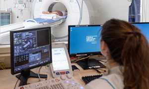
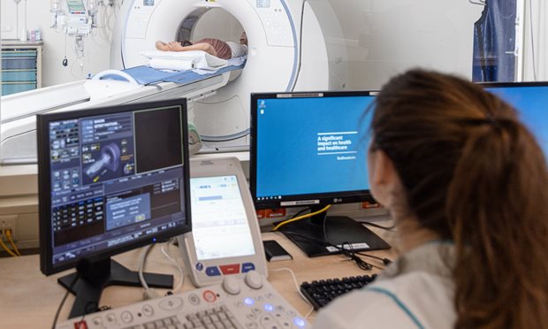
Radboudumc Technology Center Imaging
This technology center provides cutting-edge technology and service for imaging-related preclinical and clinical research questions.
see pageDosimetry Core Unit
The goal of the Dosimetry Core Unit (DCU) is to support effective and safe radionuclide therapy for cancer patients. To this aim, we perform translational research, covering the whole range from bench to bedside to community.
see page