About our equipment
Optical imaging offers unique possibilities for in vitro and in vivo imaging applications, especially in the context of molecular imaging.
read moreApplications in PRIME
The MILabs optical imaging module and the IVIS Lumina can be used for the following applications:
read moreApplications in PRIME
The MILabs optical imaging module and the IVIS Lumina in PRIME can be used for the following applications:
- To study the physiological, pathological and molecular processes in real time and longitudinally;
- For diagnostics, pharmacokinetics, pharmacodynamics, and drug screening in preclinical studies.
This imaging modules are used in PRIME for:
- Tumor imaging with fluorescence imaging
- Skin infection models
- Wound healing
- Malaria infection/immunization
- Disease-inducible promoters for inflammation
- Rheumatoid arthritis and osteoarthritis models
IVIS Lumina Caliber
Standard fluorescent filter sets
|
Filter set |
Label |
Background Passband (nm) |
Excitation Passband (nm) |
Emission Passband (nm) |
Dyes, Fluorescent proteins, and Quantum dots |
|
1 |
Green |
410-440 |
445-490 |
515-575 |
GFP, EGFP, FITC |
|
2 |
Red |
460-490 |
500-550 |
575-650 |
DsRed, PKH26, Qdot® 605 |
|
3 |
Far-red |
580-610 |
615-665 |
695-770 |
Cy5.5, Alexa Fluor, Qdot®705 |
|
4 |
NIR |
665-695 |
705-780 |
810-885 |
ICG, Qdot® 800 |

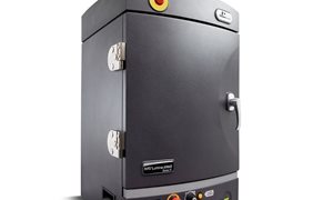
Optical Imaging unit MILabs
The MILabs OI units can be used for bioluminescence, fluorescence and Cherenkov imaging, and it can even be combined with the MILabs U-SPECT-6/CT at our facility, to do multimodal imaging. Researchers at PRIME use it for a wide variety of applications to answer their research questions, such as ex vivo imaging of luminescent cells seeded in bone disks, in vivo follow-up of tumor growth through luminescence imaging, or in vivo biodistribution of fluorescently labeled compounds.