What is an MRI examination?
With an MRI scan potential abnormalities of the prostate gland can be made visible. The information provides only a general description of a procedure. It may be that your specialist requests a procedure that may vary from the one described here.MRI Checklist
Please read and complete the following checklist before your appointment. download the pdf fileThe examination
Before the examination
Because we are examining your prostate, some additional preparations should be made. lees meer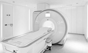
During the examination
When you check in at the Radiology Department, your details will be verified and corrected if necessary. Should you have forgotten to bring your ´check list´ then you will be required to fill out a new questionnaire. read moreDuring the examination
You are required to report to the Radiology Department (Route 780) 10 minutes before the time stated on your appointment card or letter, keeping in mind that it’s a 10 minute walk from the main entrance to the Radiology Department.
The radiographer or assistant will collect you (and your companion) from the waiting area, bring you to a changing room where you (and your companion) will be required to leave all metal objects, telephones, credit-bank cards etc, behind. An intravenous line will be inserted and the procedure will be explained. You may
direct any questions to the radiographer at any time before or during the preparation for your examination.
Buscopan
In all cases it is necessary to administer a medication, Buscopan®, to reduce movement of the bowels and so decrease motion artefacts on the images. This is administered by the radiographer via the intravenous line, just before the start of the examination. You may not receive these medications should you have:
- Increased pressure in your eyes (glaucoma)
- Increased or irregular heart rhythm
- A muscular disease
- Stricture of the colon (large bowel)
- A problem with urination whereby you have to insert a catheter to empty the bladder.
Should you have any queries over this medication you may contact your specialist, radiology department or ask the radiographer during the preparation for your examination.
Contrast Agents
It is possible that a contrast agent may be administered during your scan. This is determined by the specialist and radiologist and allows the differences between organs and tissues on an MRI scan to be seen more clearly. To administer the contrast agent the radiographer will insert a cannula/intravenous line and will remove the cannula at the end of the examination. During the examination the radiographer will administer the contrast agent when you may experience a cold sensation in the arm or a strange taste in the mouth. These sensations only last a few seconds. Rarely do patients have a reaction to the contrast agents used for MRI scans, but should you have had a reaction in the past, please contact the Radiology Department as the radiologist can in consultation with your specialist, take preventative measures. If you have a reduced kidney function we would also request that you contact the Radiology Department.
In the MRI examination room
When you enter the MRI examination room, you will be required to lie on the scanner table. To detect the tiny radio signals that are emitted from the body, a ´receiving device´ is placed behind or around the area to be examined. You will be given a rubber ball to hold onto during the scan. This is the alarm bell and the radiographer will instruct you as to when you can use for any emergency. On hearing the alarm, the radiographer will check on you and remove you from the scanner if necessary.
The part of your body being examined is placed in the middle of the scanner (an open-ended, cylinder-shaped machine about a one and a half metres long). During the examination you will hear different kinds of loud noises (knocking/buzzing) but will be given earplugs or headphones to wear. If you wish you may listen to the radio. During the examination many images are taken. Some images only take a few seconds to make, others several minutes. Once the noise has stopped, then one set of images have been made. During the making of the images and the time in between, you will be required to lay very still. Once all the images have been made the radiographer will slide you out of the scanner and remove the intravenous line. The whole examination takes on average, around 30 minutes, but in some cases can take up to an hour or longer.
Results PI-RADS
Your prostate MR images are evaluated by a specialized radiologist using the PI-RADS score. read moreResults PI-RADS
Your prostate MR images are evaluated by a specialized radiologist using the PI-RADS score. With this assessment, the radiologist determines the likelihood of the presence of prostate cancer. The PI-RADS score is classified in a categories form 1 to 5.
Overview of PI-RADS scores (the likelihood of prostate cancer):
- very low (very unlikely)
- low (unlikely)
- intermediate (unclear)
- high (probably present)
- very high (very likely present)
Your practitioner will discuss the results of the MRI with you during your next appointment.
More information
Getting there
Visiting address
Radboudumc main entrance
Geert Grooteplein Zuid 10
6525 GA Nijmegen
Directions


Changing your appointment
Would you like to change or cancel your appointment? You can indicate this in your online personal medical file ’mijnRadboud’ (only in Dutch) or contact the department where you have an appointment. We will schedule a new appointment with you as soon as possible. login mijnRadboud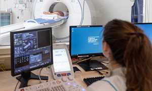
Department Medical imaging
The department of medical imaging examines and treats various conditions, and has three focus areas: anatomy, nuclear medicine and radiology.
go to page