
Research at Radboud university medical center
We create scientific impact that leads to innovation in the health and healthcare of the future.
The biology of tumors is studied at the macroscopic (PET) and microscopic (cell, subcellular) level. The aim is to compare different functional imaging modalities for the same tumor.
The focus is on:
The backbone of this system are:
This system can be extended by:
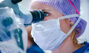
At the microscopic level, the tumor biology is studied in a quantitative manner with preservation of the tissue architecture and spatial associations. Therefore, we have developed a system for co-registration and quantitative analysis of micro-environmental phenotypic tumor characteristics. The method is based on immunehistochemical detection of multiple fluorescent signals in complete tissue sections.
Components of our digital imaging systems:
Our laboratory has permission for genetically modified organisms ('ML-II').
The facilities are:
Information will be given shortly.
We create scientific impact that leads to innovation in the health and healthcare of the future.


