About our equipment
High Frequency Ultrasound and Photoacoustic Imaging is a technique used to visualize detailed internal structures and function with unprecedented temporal and spatial resolution. read moreAbout our equipment
While ultrasound imaging is a well-established modality for clinical and pre-clinical applications, photoacoustics, on the other hand, is a young optical technique that recently gained a tremendous interest in the biomedical imaging field. In PA a short pulse of light is used to irradiate the region of interest. The instantaneous absorption of energy by naturally occurring chromophores such as blood, or exogenous injected dyes and nanoparticles induces a localized short temperature rise, which results in the generation of an US pulse via the thermoelastic effect. Using US detector placed outside the medium, the generated acoustic waves can be detected and by means of image reconstruction, the location of the US sources can be determined to allow for a two or three-dimensional visualization of the absorber distribution.Unlike the commonly used optical techniques, such as diffuse optical tomography, optical coherence tomography and fluorescence microscopy which suffer either from poor resolution or limited penetration depth due to the light scattering phenomena, photoacoustics combines high penetration depth and sub-millimeter ultrasound resolution, thanks to the weak ultrasonic scattering in biological soft tissues.
Photoacoustic imaging has the potential to provide real-time, non-invasive functional imaging of numerous prevalent diseases including tumor detection, cerebral imaging, burn wounds, pharmacokinetics, cardiovascular dynamics, rheumatology and therapy monitoring
Applications
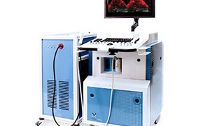
The Vevo 2100 is a major upgrade for PRIME’s small animal imaging capabilities. It provides high resolution, real-time, live animal imaging for a variety of studies. In particular, tumor angiogenesis detection, oxygen monitoring, cerebral imaging, pharmacokinetics, rheumatology, therapy monitoring.Equipment
- Vevo 2100 high-frequency ultrasound scanner
- Vevo LAZR photoacoustic system
- MS250: 24 MHz MicroScan transducer
- MS550D: 55 MHz MicroScan transducer
- MS700: 70 MHz MicroScan transducer
- LZ-250: 25 MHz photoacoustic probe
- Small animal physiological monitoring
Service
- High frequency (24-70 MHz) ultrasound imaging
- 3D imaging
- M-mode imaging
- Color Doppler imaging
- Non-linear ultrasound contrast imaging
- Contrast quantification analysis
- High frequency (25 MHz) photoacoustic imaging
- Oxygenation-hemoglobin analysis
- Photoacoustic signal quantification analysis
- Radio frequency (RF) data analysis