Teased fibres knopen

Afdelingen Pathologie Beelden Pathologie

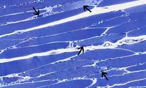
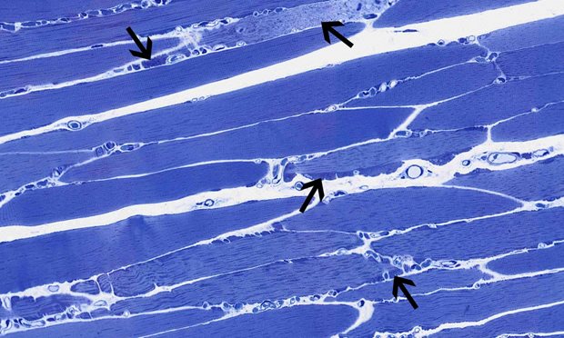
The following electron microscopical (EM) photos show some common and less common ultrastructural alterations of muscle and nerve. It may help those who practice daily muscle and nerve diagnostics, but may also aid others to learn more about muscle and nerve EM. Copies of pictures can be used for non-commercial purposes if the source is indicated.
 

Beelden

Hier staat een overzicht van alle beelden

Spier


Afwijkende mito's 1


Afwijkende mito's 2


Afwijkende mito's 3


Concentric lamellated bodies


Curvilineair bodies


Cyto­plasmatische lichaampjes


Filamenteuze lichaampjes


Glycogenose-1


Glycogenose-2


Grods-CLN10 bewezen


Grods-CLN2 bewezen


Kern inclusies


Myeline figuren


Myofibrillaire myopathie-1


Myofibrillaire myopathie-2


NCL-Curvilineaire lichaampjes


Nemaline rod-1


Nemaline rod-2


Redundant basaal membraan


Rimmed vacuolen


Semidun coupes abnormale mitochondriae


TRS tubuloreticulaire structuren


Tubulaire aggregaten


Vetstapeling


Zebra lichaampjes-1


Zebra lichaampjes-2

Zenuw


Abnormale Axons


Amyloid


Bungerse banden


Cluster sheet


Collageen pockets


Curvilinear bodies


Degeneratie


Focal folded myelin


Gedemyeliniseerde axonen


Geremyeliniseerde axonen


Gezwollen structuren met fingerprints


GRODS in ­ zenuw weefsel


Knoop van Ranvier-1


Knoop van Ranvier-2


Lafora body


Mixed­ Fingerprints-Curvilineair bodies


Ongemyeliniseerde axonen


Onion bulb- basale lamina


Onion bulb Schwanncell-layer


Remak bundle


Renault lichaampje


Schmidt-Lantermanse insnoering


Teased fibres


Teased fibres tomacula


Tomacula


Vater Pacini corpuscle

  • Medewerkers
  • Intranet